Extending Fluorescence Quantification to Antimicrobial Material Research
2026-06-03

EzCube Green-Channel PI Fluorescence Readout in the Study of PGAIC Coatings
Application Overview
Benchtop fluorometers are widely used for precise DNA and RNA quantification. Yet in research workflows that rely on fluorescent dye signals, the same type of instrument can also support applications beyond routine nucleic acid measurement.
In a study published in Journal of Coatings Technology and Research, researchers from Ehime University and Kochi University investigated a new antimicrobial coating material, poly-γ-glutamate ion-complex (PGAIC). The study evaluated the material’s protective performance against multiple pathogens, including SARS-CoV-2, Serratia bacteria, and dermatophytes.
To better understand how the coating affected microbial cells, the researchers combined conventional colony-forming unit (CFU) counting with propidium iodide (PI) fluorescence analysis. Within this workflow, the EzCube Fluorometer was used to measure PI fluorescence intensity and support the estimation of lethal-cell changes associated with compromised membrane integrity.

Source article: Onari and Ashiuchi reported the development and application of PGAIC as an ultrabroad-spectrum microbicidal coating in Journal of Coatings Technology and Research.
Table of contents
- Why CFU Counting Alone Was Not Enough
- How EzCube Was Used in the Lethal Cells Assay
- Converting PI Fluorescence into Estimated Lethal-Cell Counts
- What the Fluorescence Data Revealed About the PGAIC Coating Mechanism
- Workflow for Microbiology and Molecular Biology Labs
- Conclusion
- Study Method Snapshot
- What Can We Learn from This Study?
- References
Why CFU Counting Alone Was Not Enough
In antimicrobial material development, viable cell counts provide only part of the picture. CFU counting helps determine whether cells remain capable of growth after exposure to a material, but it does not fully describe how membrane damage develops over time.
For this reason, the researchers paired CFU-based evaluation with PI fluorescence analysis.
- CFU counting was used to assess what the paper refers to as survival potential—whether the tested pathogens could still grow and form colonies after contact with the coating.
- PI fluorescence analysis provided another layer of information. PI can enter cells with compromised membrane integrity, allowing researchers to estimate lethal-cell counts and analyze lethal rates based on fluorescence signal changes.
Together, the two methods helped the researchers compare the loss of viable cells with the increase in lethal-cell signals, which was important for interpreting the coating’s microbicidal behavior.
How EzCube Was Used in the Lethal Cells Assay
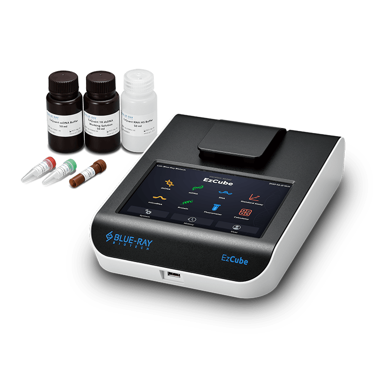
The study describes a “Lethal cells assay on PI fluorescence” in the Materials and Methods section. In this assay, S. marcescens cells were treated, mixed with PI reagent, and incubated at 25 °C for 5 minutes. Fluorescence intensity was then measured using the EzCube Fluorometer.
The paper specifies the optical settings as:
- Excitation: 490–535 nm
- Emission: 564–650 nm
- Readout: Relative fluorescence units (RFU)
In other words, the assay used EzCube’s green excitation range, paired with emission collection at 564–650 nm, to measure PI fluorescence.
Converting PI Fluorescence into Estimated Lethal-Cell Counts
To use PI fluorescence as a quantitative readout, the research team established a calibration curve with EzCube.
The paper defines the relationship as: y = 505.6x
where y represents relative fluorescence units (RFU), and x represents estimated lethal-cell counts.
The calibration showed strong linearity across 3.0 × 10⁷ to 4.8 × 10⁸ /mL, with: R² = 0.9993.
With this calibration, PI fluorescence signals could be converted into estimated lethal-cell counts, providing a quantitative basis for interpreting the coating’s microbicidal mechanism.
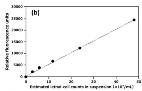
Figure: Calibration curve for PI fluorescence-based lethal-cell determination using the EzCube Fluorometer. The curve shows a strong linear relationship between RFU and estimated lethal-cell counts.
What the Fluorescence Data Revealed About the PGAIC Coating Mechanism
The study proposes a capture–killing–release cycle to explain the microbicidal behavior of PGAIC coatings.
The results suggest that bacteria were first captured on the PGAIC-coated surface. Over time, the fluorescence signal gradually increased, indicating a rise in lethal-cell signals. This pattern was not consistent with immediate cell lysis. Instead, it suggested progressive membrane damage over time.
Across the 10-hour time course, EzCube-based PI fluorescence measurements helped the researchers follow changes in lethal cells in the suspension. Combined with CFU-based survival data, these measurements provided quantitative support for interpreting the coating’s microbicidal kinetics.
Workflow for Microbiology and Molecular Biology Labs
This case highlights how fluorescence measurement can fit into broader microbiology workflows, especially when combined with complementary tools that help improve consistency in data interpretation.
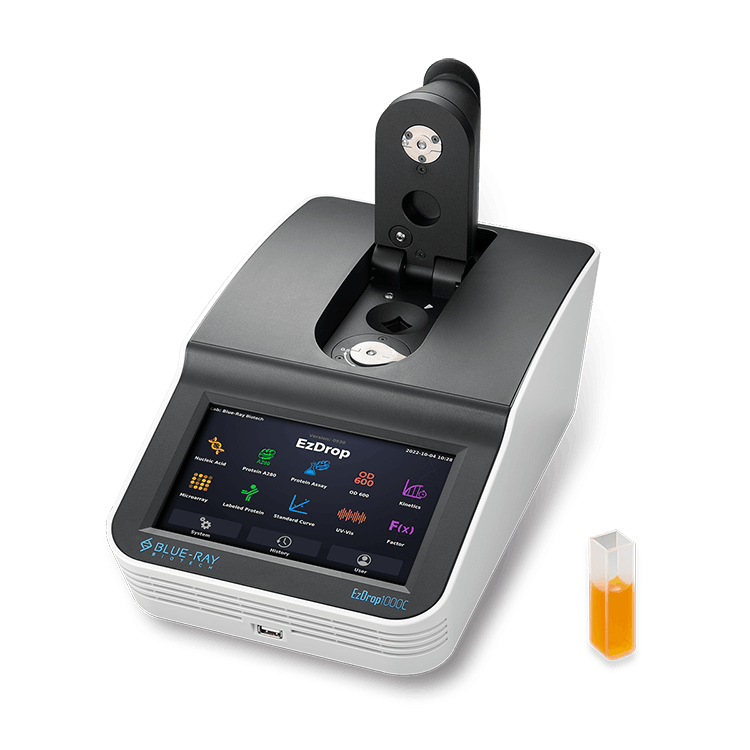
For microbial experiments, a consistent starting point is important. Before fluorescence measurement, researchers may first monitor bacterial culture concentration by measuring OD600. A spectrophotometer such as the EzDrop 1000C can be used as a complementary tool to help establish this baseline.
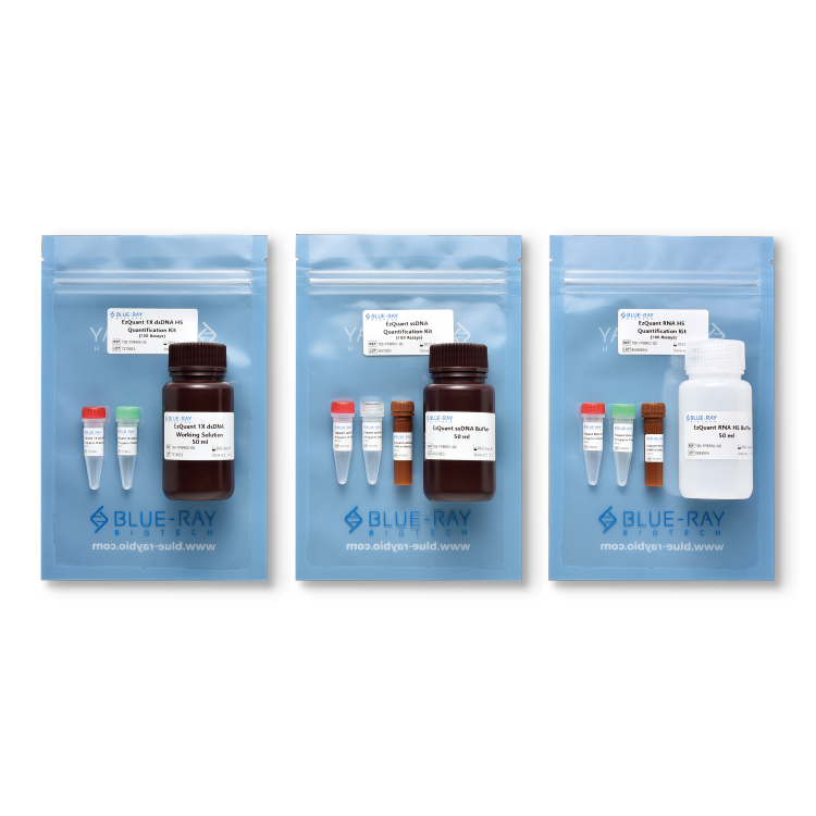
EzCube also fits naturally into routine molecular biology workflows. It offers Blue, Green, and Red fluorescence channels and fast detection within 3 seconds. When paired with EzQuant Quantification Assay Kits, EzCube supports high-sensitivity DNA/RNA quantification down to 10 pg/µL, helping researchers measure low-concentration samples before downstream workflows such as NGS and qPCR.
Conclusion
This study shows that the EzCube Fluorometer can be used beyond routine nucleic acid quantification, extending into fluorescence-based analysis for microbiology and antimicrobial material research.
Using PI fluorescence measurement under green excitation, the researchers were able to follow lethal-cell changes over time and gain additional insight into the microbicidal behavior of PGAIC coatings.
For labs working across molecular biology and microbiology, EzCube offers a practical way to add fluorescence measurement into daily workflows. It can support routine DNA/RNA quantification as well as specialized fluorescence-based assays.
Study Method Snapshot
| Item | Details from the Study |
|---|---|
| Assay | Lethal cells assay on PI fluorescence |
| Dye | Propidium Iodide (PI) |
| Instrument | EzCube Fluorometer |
| Excitation | 490–535 nm |
| Emission | 564–650 nm |
| Incubation | 25 °C for 5 minutes |
| Readout | Relative fluorescence units (RFU) |
| Calibration curve | y = 505.6x |
| Linearity range | 3.0 × 10⁷–4.8 × 10⁸ /mL |
| Linearity | R² = 0.9993 |
In this workflow, “green-channel PI fluorescence analysis” refers to PI fluorescence measurement using EzCube’s 490–535 nm excitation range and 564–650 nm emission range.
What Can We Learn from This Study?
1. Is EzCube limited to DNA/RNA quantification?
No. In this study, EzCube was used for PI fluorescence measurement in a lethal cells assay, showing a practical application beyond routine nucleic acid quantification.
2. Which EzCube fluorescence setting was used?
The paper used EzCube under excitation at 490–535 nm, with emission collected at 564–650 nm, to measure PI fluorescence intensity.
3. Why combine CFU counting with PI fluorescence analysis?
CFU counting evaluated survival potential, while PI fluorescence analysis helped estimate lethal-cell changes associated with compromised membrane integrity. Together, the two approaches provided a broader view of the PGAIC coating’s microbicidal behavior.
References
- Onari, T., & Ashiuchi, M. (2026). Rapid formulation and application of poly-γ-glutamate into an ultrabroad-spectrum, safer microbicidal coating. Journal of Coatings Technology and Research. DOI: 10.1007/s11998-025-01239-9
- EzCube Fluorometer
- EzQuant Quantification Assay Kits
- EzDrop 1000C Spectrophotometer

Reviewer
Jeffrey Lai
Senior Product Manager, Blue-Ray Biotech
Jeffrey brings over 30 years of life science experience to his role at Blue-Ray Biotech. He previously led technical support for a top-tier US PCR brand and a major laboratory distributor in Taiwan, working closely with countless researchers to solve complex assay challenges. At Blue-Ray Biotech, Jeffrey uses these field-tested insights to design products that are both innovative and practical. Off-duty, he is an avid rider who enjoys touring Taiwan’s scenic highways on his heavy bike.